New Patients Welcome
Technology
State-of-the-Art Dental Technology at Our Practice
To ensure you get the best results possible from your treatment, Dr. Heather Martinson has equipped her dental office with the latest technology, including:
Cone Beam CT
A CBCT is an advanced imaging device that takes a complete 3D picture of your mouth and sinus cavity. Unlike X-rays, a CBCT scan produces a higher-quality image that can see both hard and soft tissues in your oral cavity. Plus, it uses 10x less radiation than X-rays. CBCT scans also allow Dr. Martinson to effectively practice the BaleDoneen method of heart attack and stroke prevention.
CEREC Machine
Usually when you get a dental crown, the dentist has to send a mold out to a laboratory to create the finished product. This means you have to wait days or even weeks to have your smile fully restored. But our Chairside Economical Restoration of Esthetic Ceramics (CEREC) machine allows Dr. Martinson to create your custom dental crowns in as little as one hour.
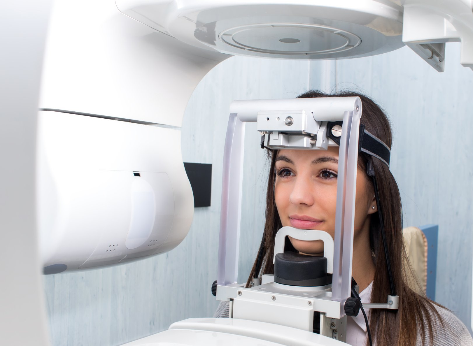
What Is A CBCT Scan?
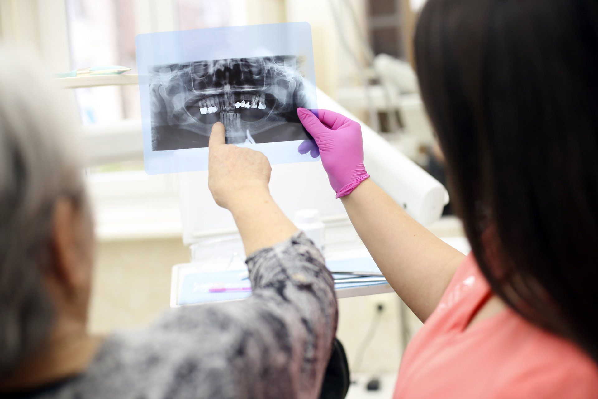
Dental Cone Beam Tomography [CBCT] is a specialized type of x-ray that provides more information than conventional dental or facial x-rays. This computerized scan uses advanced technology to generate three-dimensional[3-D] images.
What are the benefits of a Dental CBCT scan?
The benefits of a Dental CBCT scan are that it:
- Provides 3-D images of dental structures, soft tissues, nerve paths and bone which are considerably more detailed than conventional two-dimensional dental x-rays.
- Is simple and comfortable to take and can diagnostically image both bone and soft tissue simultaneously.
- Reduced radiation: Dental scanners reduce the amount of radiation the patient is exposed to, as they require little time to scan and focus on particular regions.
- Exceptional accuracy: 3D images provide far greater detail compared to using molds. Dentists are able to see pathologies, infections, nerves, musculature, and more.
- Efficiency: Scanning using 3D dental scanners is much less time consuming than traditional molding or imaging methods.
- Synergistic technologies: Data from multiple 3D scanning technologies can be combined to produce increasingly accurate models.
What are the common uses of a Dental CBCT scan?
Dental CBCT scans are commonly used to:
- Evaluate the position of teeth, bone structure, jaw joints and the airway.
- Aid in:
- Accurate placement of implants
- Surgical planning for the removal of impacted wisdom teeth
- 3-D orthodontic evaluation
- Complex root canal diagnosis and treatment
What are the risks of a Dental CBCT scan?
Although relatively low, Dental CBCT scans cause some exposure to radiation; the amount of exposure is approximately the same as taking a five-hour international plane flight. Due to radiation exposure, scans are not generally recommended for pregnant women and should be used cautiously in the pre-orthodontic evaluation of children.
How does a Dental CBCT scanning procedure work?
During the scan, a motorized arm rotates 360-degrees around your head while capturing multiple images from different angles that are then reconstructed to create a single 3-D image.
Who interprets the results of a Dental CBCT scan?
The interpretation of a Dental CBCT scan may be done by your dentist, dental specialist or radiologist.
DENTAL 3D SCANNING
Dental Scanning Technology #2: Intraoral Scanner (IOS)
Intraoral scanners (IOSs) are handheld devices used to capture direct optical impressions in dentistry. They are essentially cameras that project light onto an object such as a dental arch, creating a coordinate map used to generate a 3D surface model.
Mostly commonly, IOSs exist in the form of a “wand”, which the operator can directly insert into a patient’s mouth. They’re typically designed to be easy to use, with light, ergonomic grips and detachable tips of varying sizes.
CEREC
With our Cerec CAD/CAM technology we are able to make ceramic restorations like partial coverage or full coverage all-ceramic crowns for your teeth, in about an hour, while you wait and watch a movie. Our patients really appreciate the fact that they can have their final crown in one visit.
We no longer have to take an impression and make a model and send it to a dental lab to have your final crown made. You don’t have to wear a temporary crown any more and come back and get numb again to seat your final crown. It can now all be done in one visit! Dental Technology has come a long way!!
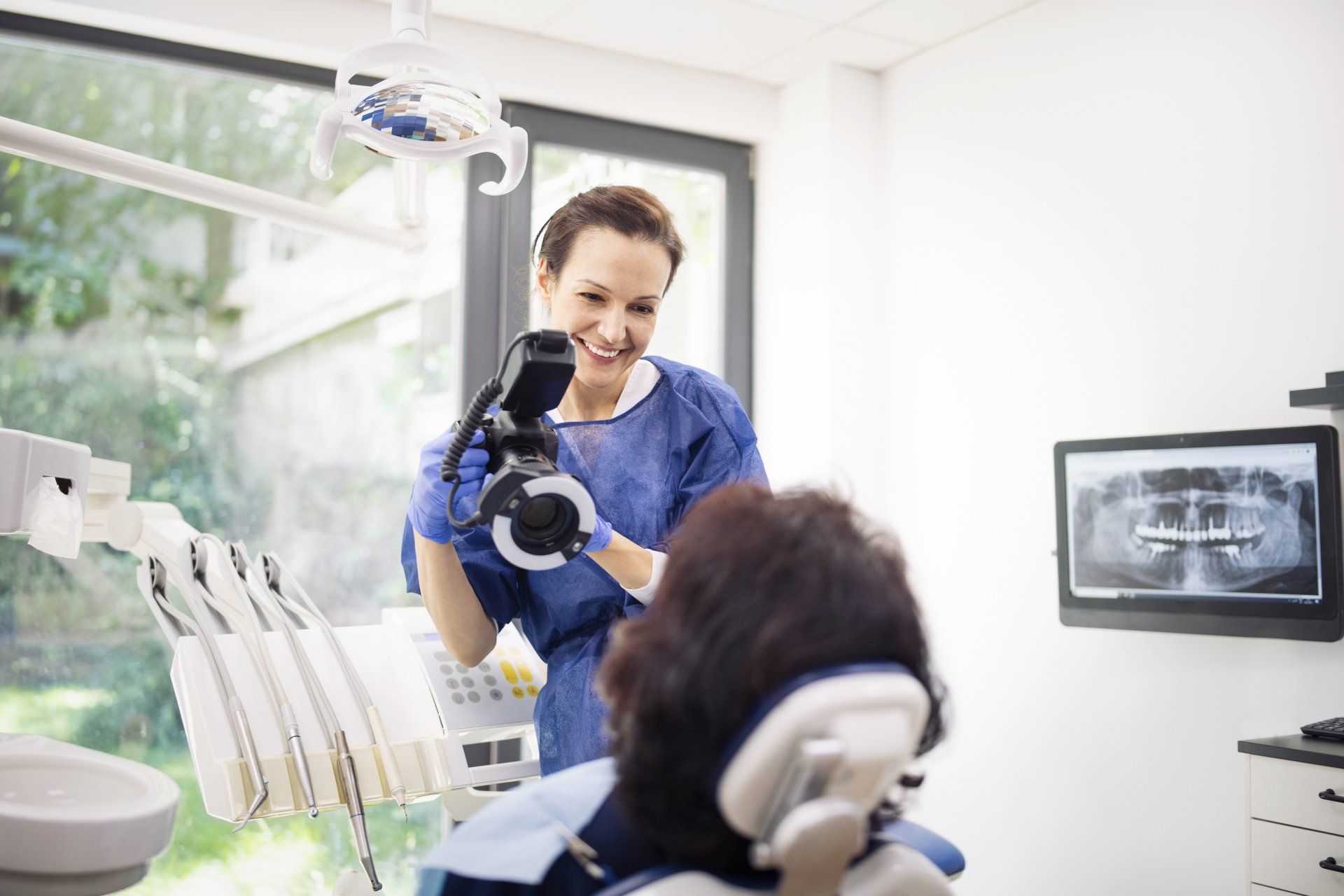
We use a digital camera to photograph your face and mouth for treatment planning purposes and to provide a visual record of your progress. In the case of restorations involving lab work, the photos are also sent to the lab to aid in crafting the highest quality restorations possible.
The Prophy Jet is an advanced cleaning tool that combines air, water and a mildly abrasive substance such as sodium bicarbonate (baking soda) to accomplish the removal of heavy stains, plaque and debris from teeth for superior teeth cleaning.
Scaling refers to the removal of stubborn calculus (hardened plaque) and bacteria from the teeth, and is commonly used in the treatment of gum disease. Ultrasonic scalers remove plaque deposits from the teeth using a minute scaling tip that vibrates at ultrasonic frequency, along with a stream of water to wash away the debris. The vibrations of the scaling tip also create a shockwave that destroys the cell membranes of the bacteria present in the plaque, effectively killing them.
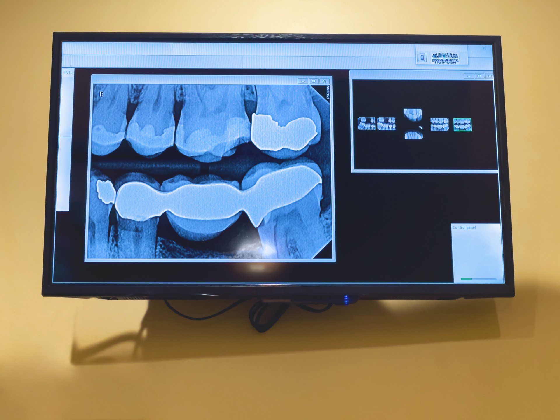
Our digital X-rays will allow you and our doctors to immediately view your X-rays on a monitor in the treatment room. Digital X-rays emit about 80% less radiation than traditional X-rays.
Panoramic X-rays produce 2D, panoramic images that give a full overview of your upper and lower jaws and teeth. Dr. Warren will use the images to evaluate your mouth in the planning for certain types of treatment.
We Save Lives And Teeth!